Characterisation and 3D structure of melanomacrophage centers in shorthorn sculpins (Myoxocephalus scorpius)
New publication by Mai Dang, Cameron Nowell, Tam Nguyen, Lis Bach, Christian Sonne, Rasmus Nørregaard, Megan Stride, Barbara Nowak

Abstract:
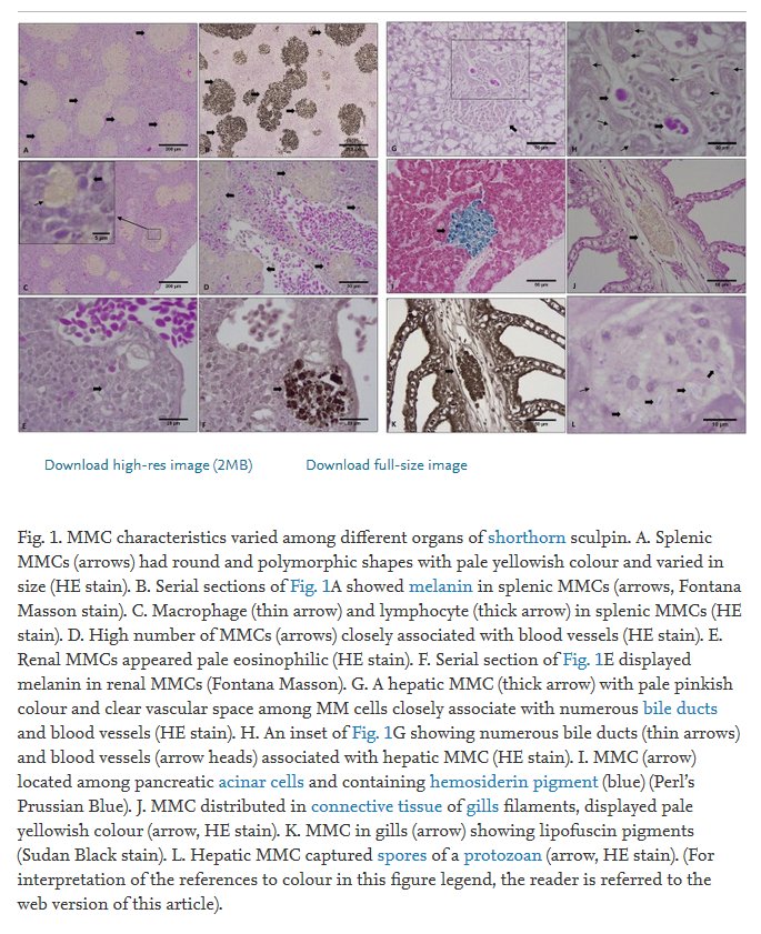
Melanomacrophage centres (MMCs) are distinct aggregations of pigment-containing cells in internal organs of fish, amphibians and reptiles. Although MMCs are commonly used as biomarkers for anthropogenic exposure in many environmental monitoring programs, a substantial knowledge on characteristics of MMCs is required prior to the assessment of MMC responses. The present study was the first to determine the 3D structure of splenic MMCs of a fish from a number of consecutive histology sections by use of the Fiji and AutoCad software. Most splenic MMCs of shorthorn sculpins (Myoxocephalus scorpius) had spherical shape and limited variation in size (maximum diameter). We confirmed the close relationship between MMCs and blood vessels in spleen of shorthorn sculpins as 97% of investigated MMCs (60 whole MMCs over 510 μm thickness of the samples) were closely associated with splenic blood capillaries (mainly ellipsoids) at least once in a set of consecutive sections. In this paper, we describe variations in morphology, density, size, area, distribution, pigments and response to pathogens of MMC populations from different organs (spleen, kidney, liver, pancreas and gills). Additionally, we provide evidence suggesting the presence and dominance of pheomelanin in MMCs of shorthorn sculpins.
